Friday, February 29, 2008

Does anyone walk less than a radiologist?

Radiology is a fairly sedentary occupation. Unlike other physicians, who spend their days scampering from patient to patient or slogging through endless hospital rounds, we sit quietly in the dark all day, staring at images. After learning of the following paper via the Dr Shock MD PhD blog, I began to wonder -- just how sedentary is my specialty? This seemed like a fine topic to research for a Leap Day post.
How many steps does a doctor take in the hospital? No difference between internists and general surgeons, but a relationship with age and BMI. Goosen JHM, Ettema HB, Kollen BJ, Verheyen CCPM.  Ned Tijdschr Geneeskd. 2008;152:203-6.
Disclaimer: The original paper is in Dutch, and my Dutch skills are on a par with my Croatian, Chinese and Tagalog skills. Therefore, I turned to good old Google Translate for assistance in reading this article.
These researchers tagged a group of 131 internists and 131 surgeons with pedometers. The internists walked an average of 5457 steps/day (564 steps/hour), while the surgeons walked an average of 5193 steps/day (536 steps/ hour). This difference was not statistically significantly different.

To put this in perspective, the authors compared these physicians with the hourly step rates of air crew serving on long haul flights (842 steps/hour) and of patients with prosthetic (artificial) hip joints (143 steps/hour).

As it turns out, I just happen to wear a pedometer every day to keep track of my own activity (or lack thereof). Below is a plot of my daily step count, which averaged 4909 steps/day for the past week.


The spikes of activity on days 2 and 5 are, respectively, days in which I went folk dancing and spent some time walking on a treadmill. After eliminating these two outliers, my average drops to 3108 steps/day, or 194 steps/hour (during the 9 work hours out of 16 hours awake). This is probably a pretty fair estimate of my step rate while at work. Plotting this up against the other groups, I got the following plot:


Is my step count representative of all radiologists? I don't know, but I suspect I'm probably somewhere in the middle of the pack.


So, I can finally answer the question posed by the title of this post: does anyone walk less than a radiologist? The answer is yes, patients with hip prostheses walk less than a radiologist (at least this one). Can it get any more pathetic than that?

Short of pathologists or paraplegics, I don't see how. I guess this would be a fine time for me to start walking laps of our building during lunch.

Thursday, February 28, 2008

Skeptics Circle - 81st edition

The Skeptics' Circle is a biweekly carnival for bloggers who apply critical thought to questionable stories.

The latest  (81st) edition is just out on the Conspiracy Factory site, hosted by Factician.

No paltry listing of blog links this -- this episode takes a few days after the lunar eclipse, where
You're hidden in the bushes, in the dark. Peeking over the hill in front of you, you can see a small group forming at the base of the hill. It's a group of men and women in black suits, carrying torches, and they're filing towards what looks like a circle of barrow mounds....
My contribution to the current conspiracy is posted adjacent to the "Finish Him!!" graphic.

Wednesday, February 27, 2008

Take the First Chernoff on the Left

There is no topic, no matter how intrinsically tedious or boring, that cannot be made even more tedious and boring with the right set of Bad Powerpoint Slides.
The converse is also true -- a few engaging images can make even a fairly mundane topic sing and dance. Radiology lectures are an obvious exemplar of this, being filled with lots of fascinating images. However, what if you need to present a ton of data? If so, beware -- your task will be a mighty one. Decades of punitive Powerpoints have pretty well proven that any slide with a big table of data is going to have an LD50 of under 30 seconds.

A lot of creativity has gone into solving this problem. One intriguing method was proposed by Herman Chernoff back in 1973 in his paper: "The Use of Faces to Represent Points in k-Dimensional Space Graphically", Journal of the American Statistical Association 1973;68:361-368.

Chernoff's proposal relies on the notion that the human eye-brain combination is one of the most powerful pattern-recognition engines on the planet. If one is trying to convey a large multidimensional table of numbers, he suggested representing each observation as a computer-drawn cartoon of a human face,
...whose features, such as length of nose and curvature of mouth, correspond to components of the point. Thus every multivariate observation is visualized as a computer-drawn face. This presentation makes it easy for the human mind to grasp many of the essential regularities and irregularities present in the data.
For example, consider the following table of totally bogus data that I made up just now, all by myself. It purports to show income level, quality of life and onerousness of call for 4 physician specialties.

  Income Quality of Life Onerousness of Call
Radiology Medium High Medium
Pediatrics Low Medium Low
Family Practice Low Medium High
Surgery High Low High

Yep, this table is a real dozer, all right. But wait! What if we represent the same data as Chernoff faces? To do this, I fired up my copy of Mathematica and used it to run some sample code I downloaded from the Mathworld site (R enthusiasts see here for sample code). In the plot below, the shape of the eyes, mouth and head respectively represent income, quality of life and onerousness of call. My fabricated data now looks something like this:

Of course, this kind of plot has certain potential flaws. As K. S. Park points out:
A major drawback of Chernoff faces is that the subjective assignment of facial expressions to variables affects on the shape of the face.
No feces, Conan-Doyle character. My fabricated and highly biased data, coupled with an equally biased assignment of facial features, makes that first Chernoff on the left look pretty darned appealing, doesn't it?

A discussion of Chernoff faces wouldn't be complete mentioning the work of Eugene Turner, particularly his prize-winning map titled Life in Los Angeles. He has this to say about it:
I compiled and designed this map which was drafted by Richard Doss. It was an idea based on Chernoff faces. It is probably one of the most interesting maps I've created because the expressions evoke an emotional association with the data. Some people don't like that.
Here's one final fun example from the world of baseball: Alex Reisner's What's the Matter with Chernoff Faces, illustrated by his plot of the 2005 National League season. I especially enjoyed the alternative system he suggests there: Reisner faces.


Update, 2/28/08:   I realize that I sort of left things hanging here, with no final socko conclusion rendered on the utility of Chernoff faces.  This is probably because I still don't have a strong opinion.  However, now that I have the machinery to plot these faces, I'll look for ways to apply them to real data and see how they work for me.  My goal is to avoid using them like the person in Andrew Lang's quote, who
...uses statistics as a drunken man uses lamp-posts -- for support rather than illumination.

Tuesday, February 26, 2008

Grand Rounds - Vol 4, No. 23

The latest Grand Rounds (issue 4.23) is up on ScienceRoll!  

Grand Rounds is a moveable feast that features some of the more interesting posts from the  past week in the medical blogosphere.

My contribution for this edition describes a recent dinner I attended, where the conversation suddenly turned to skulls...  

Pareido-Leo

Q. What's the definition of an experimental drug?

A. Anything you inject into a rat that generates a scientific paper.

A zillion years ago, I did my subspecialty training on the very first clinical MR machine on the whole West Coast. Heck, for the best part of a year, we had the only clinical MR machine on the whole West Coast. Having this machine gave us special god-like powers over the researchers at every other med school within 1500 miles. MR was such a hot-button topic in those early days that it was relatively easy to get MR papers through peer-review. It seemed as if one could throw just about any body part into an MR scanner, collect a few similar cases, and some radiology journal somewhere was hungry enough to publish the paper about it.

There was so much competition for this single scanner that each section of the radiology department got 2 scan hours / per week to use for their research. Just 2 hours meant just 2 patient scans, so every readout session became a big deal for us. Our whole section would troupe together down to the MR room twice a week to read out our single MR case for the day.

There was a clear hierarchy for these sessions: the Great Professor would sit in the comfy chair in front, and hold forth on the findings. The lesser professors would sit a bit further back. The fellows, residents, medical students and visiting foreign fellows were spaced around the lightbox according to their station in the food chain. Sometimes the Great Professor would ask a lesser being to describe the findings -- a sphincter-clenching moment for many.


One day we were looking at a pelvic MR image just like the one above, and the Great Professor asked us what we saw. A great peristaltic wave of silent clenching spread through the crowd, because we lesser beings all saw bubkes. We watched with bated breath as the Great Professor continued to gaze at the image, waiting to learn about the subtle finding that we had all missed. Finally, he said, "Looks just like a lion's face.", got up and walked away.

This was not my first experience with radiological pareidolia, but it remains one of the more stressful. In teaching conferences nowadays, I sit in the comfy chair on the front row. It's a dirty job, but I've taken up the torch from the Great Professor and periodically show my residents and fellows cases just like the one above.

Good times.

Monday, February 25, 2008

Graph Fu

See this stunning Flash-based plot on the NY Times site, showing movie box office receipts over time. (via Daring Fireball)

Giving presentations is a big part of the life-blood of academic radiology. Therefore, I'm always looking for cooler and more effective ways of presenting information.

After seeing this lovely Times plot, I'll have to see if I can't convince R, my favorite stat environment, to make some similar plots of my data.
-----------

Update:  For those geeky enough to care, someone else has already worked out how to make R do a very similar plot.

Sunday, February 24, 2008

Using X-rays to Sell Shoes

Consider this text from a radio commercial circa the late 1940's.
Every parent will want to hear this important news! Now, at last, you can be certain that your children's foot health is not being jeopardized by improperly fitting shoes.
Wow! How could any parent resist this call to action? If you're not convinced, consider this quote by J.J. Lowe:
With this apparatus in his shop a shoe merchant can positively assure his customers that they never need wear ill-fitting boots and shoes; that parents can visually assure themselves as to whether they are buying shoes for their boys and girls which will not injure and deform the sensitive bone joints.
Fortunately, there's no need for alarm. The product being hawked in the quotes above -- a shoe-fitting fluoroscope -- is no longer used.

The idea of using X-ray technology to improve the fit of shoes probably arose during World War I. Shoe-fitting fluoroscopes were patented in the U.S. and the U.K. in the 1920's and were common fixtures in shoe stores throughout North America and Europe from the late 1920's through the 1950's.

In an excellent review of this topic, Duffin and Hayter state:
In our opinion, however, the shoe-fitting fluoroscope was nothing more nor less than an elaborate form of advertising designed to sell shoes. It entered a well-established culture of shoe-selling hucksterism that relied on scientific rhetoric; it took advantage of the woman client newly accustomed to the electrification of her home and the patter of experts' advice about "scientific motherhood"; it neatly sidestepped the thorny problem of truth in advertising that became an issue in the interwar years; and it enticed thrill-seeking children into shops where salesmen could work their magic.

I can still remember looking at my 6 year-old feet through one of these devices in my local Sears store. It was extremely cool to watch my toes wiggle on the green glowing screen, and I was very disappointed when the machine was eventually removed from the store. However, I felt much differently about these devices once I became a radiologist and began to learn more about radiation and its effects on the body.

Physicians use fluoroscopy to perform all sorts of patient procedures ranging from barium enemas to cardiac catheterizations to the placement of prosthetic joints. However, we only expose our patients to the minimum amount of radiation necessary and we keep ourselves way the heck out of the way of the X-ray beam.

By contrast, shoe-fitting fluoroscopes were badly designed, meant to be used by barely-trained shoe salesmen, and gave a considerable dose of radiation to one's feet and other body parts. A customer using this machine would be essentially standing on the the X-ray tube, separated from it only by 1 mm of aluminum shielding. Body parts in the main X-ray beam (customer's feet and salesman's hands) received most of the dose.

But wait -- there's more! These X-rays not only went up through the feet to the fluoroscopy screen, but continued upwards, through the heads, thyroids and eyes of the users. Not exactly an optimal design. Even body parts not directly exposed to the main beam could receive a considerable dose from scatter radiation. As an exercise in discomfort, look at the diagram below and visualize just what sort of dose your own personal gonads would get while standing on this thing.

Drawing after Bushong SC and West WD. Exposure from a Shoe-Fitting Fluoroscope. Health Physics 1970;18:575-576.

Duffin and Hayter state:
In addition to the dose received by the feet, the entire body of the customer -- along with her parent and the attending salesman -- was bathed in radiation; others in the shop were also being irradiated through the walls of the machine. In the waiting-room chairs, the permissible daily dose could be received by a single person in one hour.
It's therefore ironic that these machines were targeted at two of the more radiation-sensitive segments of the customer base: children and women in their child-bearing years. The following quotation is excerpted from the installation instructions of one machine:
Of course, it should face the ladies' and children's departments by virtue of the heavier sales in these departments.

With the machines of the time, a child could be exposed to 10 to 116 R in a 20-second period, a dose sufficient to cause radiation burns to the skin. If one considers further that a given child might try on several pairs of shoes per visit with several visits per year, these multiple exposures could add up to an even higher yearly dosage. To put this in perspective, the daily permissible dose for radiation workers in 1946 was 0.1 R.

By the way, the question, "Dude, what's my radiation dose?" is not unlike another question: "Dude, how much does it cost to fly to Chicago?". The answer to both questions is, "It depends." I've paid anywhere from $300 to $1500 to get to Chicago, depending on factors such as how long before the flight I booked my ticket, coach vs. first class, one-way vs. round-trip ticket, and my length of layover. The effective radiation dose depends upon a similar number of variables, such the type of radiation, the length of the exposure and just which body part is exposed. Exposure to critical tissues such as the eye, thyroid and gonads is a much bigger deal than exposure to a relatively radiation-insensitive area like the foot.

Were the medical experts of the day asleep at the wheel while all of this was going on? Not really. The British X-Ray and Radium Committee of Great Britain issued recommendations for workplace radiation dosage in 1921, and Americans followed suit in 1922. However, these machines remained unregulated until 1948, when New York became one of the first jurisdictions to regulate their use. The American College of Radiology published a warning editorial in the Journal of the American Medical Association in April 1949. Its conclusion:
The bitter fact remains that fluoroscopy simply cannot be really safe in the hands of those untrained to its use and relatively ignorant of its dangers.
This was also a time of growing post-war public concerns about radiation safety, likely related to WWII events such as the Manhattan Project and the atomic bombing of Japan. Pennsylvania became first state to ban these shoe-fitting fluoroscopes in 1957. Thirty-three other states followed suit with some form of legislative action. In 1963, these devices were finally banned in the U.S.

Has anyone ever actually been harmed by shoe store fluoroscopy? Alas, only anecdotal evidence is available to help answer this question. In 1957, Kopp described a case of a shoe saleswoman who developed chronic radiation dermatitis on her own feet. More recent reports of basal cell carcinoma of the foot attributed to shoe store fluoroscopy have been reported by Oster-Schmidt in 2002 and Smullen in 2007.

From what we know of radiation exposure in animal experiments and from studies of atom bomb survivors and nuclear industry workers, a number of other problems can be hypothesized: foot-bone deformities in child customers, and testicular tumors and leukemia in salespeople. Unfortunately, due to the undocumented dosage and time course of the exposure, and to the long lag period between radiation and some of its side effects (e.g. cancer), the long term health consequences of shoe store fluoroscopy will likely never be known.

Although shoe-fitting fluoroscopes are no longer used, their story remains a cautionary tale in the ongoing struggle against quack medicine. The storyline of shoe-store fluoroscopy follows an arc familiar to woo-watchers:
  1. a real physical phenomenon or technology is discovered
  2. unsubstantiated health claims are made about it
  3. the potential dangers are discounted
  4. quack usage becomes popular and spreads widely
  5. government regulation catches up slowly, if not circumvented politically
However, unlike forms of woo which are essentially placebos (e.g. homeopathy and acupuncture), shoe-fitting fluoroscopy was based on a phenomenon whose effects could actually be objectively measured: ionizing radiation. The scientific research necessary to demonstrate the harmful effects of such radiation took decades to accumulate, but eventually became overwhelming enough to convince even politicians and regulators to do the right thing, just as the fad was passing.

Acknowledgements:

The images of the fluoroscope and the shoe-fitting certificate are used with the kind permission of Dr. Paul Frame, of the Oak Ridge Associated Universities Museum.

Many of the historical details for this post were culled from three excellent resources.  I highly recommend reading the originals:

1) the online exhibit on shoe-fitting fluoroscopy at the ORAU Museum, gives a very nice introduction into the history of this device with some excellent illustrations.

2) Baring the Sole: The Rise and Fall of the Shoe-Fitting Fluoroscope is the definitive history of this device, and was published in 2000 by Jacalyn Duffin and Charles Hayter, Isis 2000;91(2):260-282.
3) When the solution is the problem: a brief history of the shoe fluoroscope.  Nedd CA 2nd.  AJR 1992;158:1270.

Saturday, February 23, 2008

Phone Sects

Mellophone picture from the WikipediaLast week at dinner, a friend raised the following question:
"If a native English speaker is called an anglophone and a native French speaker is called a francophone, what is the term for a native Spanish speaker?"
My immediate wise-ass reply was: "A yo-phone."

Flippant answers aside, none of us knew the answer. The question is a good one -- are there similar "-phone" words for other languages? I decided to hunt for answers in the Source of All Knowledge™, i.e. the Wikipedia. A quick trip there this evening turned up the answer to our original query -- "hispanophone". I also found a few more "phones" there:

"-phone"Usage
Allophoneany language other than English or French
AnglophoneEnglish
ArabophoneArabic
DanophoneDanish
DutchophoneDutch
FinnophoneFinnish
FrancophoneFrench
GaelophoneGaelic
GermanophoneGerman
GramophonePhonographish
GrecophoneGreek
iPhoneAppleish (term for yo-phone)
ItaliophoneItalian
LusophonePortuguese (derived from Lusitania)
MellophoneFrench (horn variant)
PersephoneHadean
RussophoneRussian
Sinophone Chinese
Swedophone Swedish
UkrainophoneUkrainian

Friday, February 22, 2008

And Then the Dinner Conversation Turned to Skulls...

Skull photo from WikipediaWhat kind of dinner was this -- a gathering of murder mystery writers? A meeting of anthropologists, paleontologists or forensic pathologists?  Ghouls?

Nope, this happened at a recent meetup of bloggers in my area. A plural of bloggers has no trouble talking forever about their blogs and recent posts. We did no less. However, toward the end of the meeting, we slid off-topic, and talked about other matters.

My table neighbor described a gift she had given to her husband: several skulls. Most husbands might not appreciate such a princely gift, but hers did. I just happen to be that kind of husband myself. I am not only fond of skulls, but also see a fair number of them at work.

Lateral radiograph of a skull
The conversation then segued to good places to buy skulls. Sweeney Todd would have no problem finding one, but where does a non-demon-barber type shop for such a thing?

As it turns out, it's actually not hard to buy a whole head at a local slaughterhouse. However, if your main interest is the skull, you will first have to remove a fair amount of squishy soft tissue (this includes brains). If you are not daunted by the thought of this, Google can be your best friend. Thus begins a very brief tangent into what it takes to clean a skull.

My first search -- "stripping bones" led me to a cute but irrlevant YouTube video that is mildly not-safe-for-work. However, this search didn't immediately yield any practical tips that I'd want to try at home.

I had much better luck with "cleaning bones", which led to several possible approaches. However, I'm not too wild about soaking a large head in a hydrogen peroxide solution, or boiling it on my stove. I'm not squeamish, but one of the many good reasons I went into radiology in the first place was to avoid having to deal with messy body parts. For the same reason, I'd rather not raise a colony of dermestid beetles at home. A piranha tank is similarly out -- besides, owning a personal pirahna appears to be illegal in my state.

If one steps back from the abyss of DIY skull maceration, one is pretty much left with outsourcing the job. If one wants to send off a bag of money and get back a skull, there seem to be no end of sites offering this service.  To see for yourself, just Google the phrase "platypus skull".

At this point, our cranial conversation moved briefly to a diamond-encrusted skull that may still be on the market for a mere $100 million. Admittedly, this looks very cool, but I may just spend my first $100 million on something else.

In our final calvarial communications, I suggested to my fellow blogger one possibility for topping her previous gift -- a way in which she could essentially give her husband his own skull for Christmas.

All she has to do is to figure out some way of getting a CT scan of her husband's head when he isn't paying attention. The scan data can then be reconstructed as a 3D picture showing just the bones. Better yet, there are actually companies out there who will take this same CT data and print it in 3D using stereophotolithography -- producing a life-like plastic skull exactly the same shape and size as her husband's.

3D Volume reconstruction of skull and neck from CT data
Hmmm, my wife always says that I'm hard to shop for at Christmas time. Maybe if I accidentally leave a copy of this post lying somewhere around the house...

Wednesday, February 20, 2008

Lunar Eclipse Tonight

The latest episode of a recurring series of total lunar eclipses airs tonight in the U.S., February 20th, 2008, Wednesday local.  The broadcast starts 0143 UTC and runs til 0510 UTC, with totality scheduled to appear onstage about 0300 UTC.

Weather in my area is threatening a picket line of clouds that will probably block any personal sightings of the event.  Therefore, I've posted a picture I took of the last one that was clearly visible at my house (8/27/07), just a bit before totality.

Duty Calls


In this latest comic, XKCD hits it out of the park yet again.

If I weren't already an XKCD fanboy, I'd be in great danger of becoming one.

Tuesday, February 19, 2008

Anatomically Incorrect

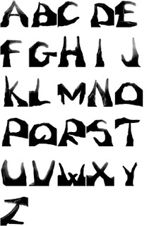
Street Anatomy has a great post on work by Björn Johansson, a very talented Swedish graphic designer and illustrator. Johansson titles this work: Anatomy of a Typeface. I, too, would love to see more of this anatomically incorrect (but wonderfully so) font.

Typeface anatomy by Björn Johansson
Anatomy and bones being central to my line of work, I was intrigued enough to click through to Johansson's website. I had a bit of trouble navigating through his site (my browser, possibly), but did find there some related work of his -- the "handwritten" font below.

Handwritten font by Björn Johansson
However, one of my favorite examples of anatomic typography is shown below.  This bit of font fu comes from the world of soccer, and involves quite different body parts. Read the hilarious setup for these posters at "Trash talking in the soccer world" by AdFreak.

Brazil vs. Argentina posters

Grand Rounds - Vol 4, No. 22

The latest issue of Grand Rounds is out on Daily Interview.  

This episode of the rotating carnival of medical blogging contains some swell postings, including musings on what actually killed Jane Austen, why nurses rather than doctors or administrators should run the hospitals, heart meds taken by the gorillas at the Cleveland zoo, and a posting from this blog, on the topic of radiology valentines

I'm happy to see that my entry easily dominated the entire category of "Are All Radiologists Geeks?".

Blog-reading helps to keep my brain from going numb while I'm working out at home.  The posts from this carnival alone should keep me conscious through several sessions.

Sunday, February 17, 2008

Desperately Seeking Supraspinatus

It's 2 am, and you need just one more wahfer-thin image of, say, the supraspinatus for a 7 am lecture to your students. Who you gonna call?

Many of us would be lying if we didn't admit going to good old Google Images at a time like this. A search I did there today for "supraspinatus MR" brought up 640 hits, which is not too shabby. However, the first 40 hits also included a bowhunting site, a dogsledding blog and a site selling Mr. Muscle Oven Cleaner. Personally, I'm going to give a bit more credence to an image I find in a peer-reviewed radiology journal than one that turns up on a dogsledding blog. I'm just funny that way.

To address this issue of search credibility, at least 2 groups of radiologists have decided to look beyond Google and roll their own radiology search engines. Of these 2, my current personal fave is GoldMiner, a site hosted by the American Roentgen Ray Society. At the instant, this engine confines its searches to a pool of 172,488 radiology images from 227 peer-reviewed journals. A search here for "supraspinatus" yielded 681 hits. Filtering with an "MRI" pull-down menu narrowed this down to 344 hits, with a listing of image thumbnails and journal citations, all linked directly to the originating journals.

Another radiologist-built search engine is Yottalook, which currently provides search services for the Radiology Society of North America. According to its website, Yottalook
is a free radiology-centric web search engine based on Google's indexing technology with proprietary relevance algorithm
This engine has been designed to search online radiology sources only, and claims to place "over 500,000 images at your fingertip". A search on Yottalook for "supraspinatus" limited for images and MRI brought up 413 hits, with a linked list of image thumbs and citations similar to that of GoldMiner. An extra feature that GoldMiner doesn't have: cute little magnified popups of each image.

IMHO, one downside of Yottalook is that it also displays a half page of Google ads for non-image searches. My non-image supraspinatus search there presented me with an ad offering "100% Guaranteed" healing of my supraspinatus at home, and linked me to an "organic" $250 home ultrasound machine. I realize that Yottalook may have little control over what Google Ads puts on their site, but the display of a goofy (i.e. exaggerated, unsubstantiated and misleading) claim like that next to my search left me with a slightly dubious taste in my mouth for an otherwise fine search engine.


One final boon for the desperate: many journal websites now include a "PowerPoint Slide for Teaching" link next to each image. Fortunately, most of us are only occasionally desperate -- and then, just enough to need one or two extra slides for our talks. However, you, too, may someday suddenly be OMG-I've-gotta-give-conference-in-2-minutes-desperate. If so, you should know that it is possible to give an hour-long case conference with only a single slide (internet access, chutzpah and an extra, non-mirrored laptop screen not included). While the students are looking at the first case on one screen, find and download the next slide on an extra screen via GoldMiner or Yottalook. Repeat PRN.
Disclaimer: The Samurai Radiologist is a member of both the ARRS and the RSNA, and has hardly, hardly ever been desperate enough to download slides in real-time while simultaneously using them a lecture.

Didgeridoo playing could improve sleep apnea

Offhand, I can't think of any connection between the didgeridoo and the practice of radiology.

I don't care.

This clinical trial by Puhan et al was conducted in Switzerland, far from this ancient instrument's origin in Australia. Somehow the authors were able to convince 25 Swiss patients with sleep apnea to play the didgeridoo. Fourteen of these 25 played the instrument for 4 months, while the 11 controls were placed on a waiting list. The bottom line:
Regular playing of a didgeridoo reduces daytime sleepiness and snoring in people with moderate obstructive sleep apnoea syndrome and also improves the sleep quality of partners

Severity of disease, expressed by the apnoea-hypopnoea index, is also substantially reduced after four months of didgeridoo playing
As a radiologist, I spend most of my work days up to my clavicles in high-tech equipment. I therefore relish all the more avocations, such as folk music and hiking, that I can enjoy with low tech gear far from the power grid. Therefore, it's a pleasing thought that a $94 plastic didgeridoo might replace a $1500 CPAP machine in some patients.


On another note, perhaps Switzerland is not such an unlikely place for this particular clinical trial at all. If anything, people who have already been playing alphorns for the past 1,800 years should be naturals at the didgeridoo.

Thursday, February 14, 2008

Radiology Valentines

Regardless of what it personally means to you, Valentine's Day is upon us again. This holiday is celebrated in a special way by those wacky radiologists. Years of training have taught them to spot tiny but clinically relevant details in a sea of noise. However, it has also made them really, really good at spotting completely irrelevant patterns. Just as normal people look for the "Man in the Moon", animal shapes in the clouds or the Virgin Mary in a grilled cheese sandwich, bored radiologists look for these sorts of things in their patients' images.

This pastime of medical pareidolia peaks at certain times of the year, particularly on February 14th and April 1st. A number of examples of this have been published in the American Journal of Roentgenology, not only one of the most respected peer-reviewed radiology journals in the world, but also one of the few with a sense of humor.

A search of the AJR archives and other radiology sites brought up a number of hits for "Valentine". If your sweetie has adventuresome tastes and you still haven't found just the right card, click through the following links for some ideas. There you will find a variety of "hearts" discovered in the midst of other organs, such as a pulmonary bulla, the bladder, Zeus only knows (some part of the gut, I think), the bladder again, the prostate, a cerebral aneurysm, the spine, the stomach, another aneurysm, a Fallopian tube, the spleen, a barium enema of the colon and other sites. One final canonical article on this topic contains no less than 27 examples of other ectopic "hearts" found by its authors.

If your taste turns to somewhat less anatomically explicit valentines, Ironic Sans has some other ideas for your scientific squeeze:
I’ve decided to honor an entirely different group of people with this collection of romantic cards you can e-mail to your loved ones on February 14th, or any other day of the year. It’s Scientist Valentines!
♥ tip for this last quote to Bad Astronomy

Tuesday, February 12, 2008

Darwin and Radiology

If radiologists imaged promiscuously up and down the animal kingdom, evolution would be a bit more obviously germane to my specialty. However, most of us don't actually image around a lot outside our own species. Instead, we spend most of our professional lives looking at one particular group of apes, which suppose themselves to already be the pinnacle of creation. How, then, can Darwin and evolution be relevant in the medical imaging of these uppity primates?

The short answer is that we visually confront the consequences of evolution daily in radiographs and other images of our patients.

When a birth defect is first discovered in a child, radiology is often one of the next steps in the diagnostic workup. Defects in one body part are often accompanied by others, and X-rays, CT, MRI and other imaging methods are used to help spot these. We've learned a lot about this clustering of anomalies by studying the development of gill arches in embryos from humans, sharks and other species. A defect in the first gill arch, for example, may result in a child with a tiny jaw and non-functioning ears.


Some body parts develop via a convoluted patchwork process that seems more worthy of the Three Stooges than any "intelligent" designer. The inferior vena cava is a great example of this. In several mammals, this large vein is formed between the sixth and eighth weeks of gestation by sequential formation, anastomoses, and regression of three paired veins. Variations in the development of these veins can lead to all sorts of wacky anomalies. Most of these venous variations are well-visualized with CT.

Another problematic legacy from the past is our spine. Our vertebral bodies are separated and cushioned by a series of discs, which are the remnant of our embryonic notochord -- a structure we share with creatures all the way back to Amphioxus. A tear in the fibrous lining of a disc can allow the gelatinous contents to extrude out of the disc, where it may cause painful pressure on spinal nerves. Neil Shubin summarized it thusly:
When we injure a disk, a very ancient part of our body plan is rupturing. Thanks a lot, Amphioxus.
On the topic of Amphioxus, I can't resist adding the following lines performed by folk musician and marine biologist Sam Hinton, who sang these words to the tune of "It's a Long Way to Tipperary":

It's a long way from Amphioxus,
It's a long way to us,
It's a long way from
Amphioxus
To the meanest human cuss.
It's good-bye, fins and gill-slits,
Welcome, lungs and hair!
It's a long, long way from
Amphioxus,
But we all came from there.
Traces of an inner fish are not only present within our patients, but within us radiologists as well. The eyes we use to view an X-ray have built-in image-processing that goes way, way back in the evolutionary tree to early sea dwellers. As I posted earlier, the human retina performs this imaging-processing before the image even reaches the brain. The edge-enhancement that occurs in the retina can help a radiologist to spot an abnormality. Unfortunately, this phenomenon can occasionally make us perceive abnormalities which actually aren't there.

So, Happy Darwin Day! I'll be spending mine ostensibly looking at human images. However, I'll also be seeing bits of a lot of other creatures today, even though I don't mention any of them on my official reports.

Sunday, February 10, 2008

There's a Spot on Your X-ray...


Not only that, but the dudes in the following video hung the chest film up backwards.

See here...

Friday, February 8, 2008

X-rays to sell jeans

What's next? Brain MR's to sell hats? PET scans to sell dog food?
“Reconstructed is the new Fall/Winter 07 print campaign for Levi’s Copper Jeans. Much the same way Copper Jeans’ seams and pockets are held in place with metal rivets, broken bones can be reconstructed with metal pins and plates. The vintage x-ray treatment is a nod to Levi’s 154 year heritage, as well helping to showcase Copper’s product features in an interesting, new light.”

Hat tip to Street Anatomy

Thursday, February 7, 2008

Image Gently

ResearchBlogging.orgIf Google led you here thinking this was a post about a lost sequel to Douglas Adams' holistic detective, turn back! Instead, it concerns a widespread movement to reduce radiation dosage in children.

A recent article by Brenner and Hall raised important concerns about safety in patients undergoing computed tomography (CT).

Brenner, D.J., Hall, E.J. (2007). Computed Tomography -- An Increasing Source of Radiation Exposure. New England Journal of Medicine, 357(22), 2277-2284. DOI: 10.1056/NEJMra072149

This paper is aimed primarily at non-radiologists. The authors give a nice review of CT, the radiation dose from CT scans, the biological effects of low doses of ionizing radiation and the cancer risks associated with CT scans.

The authors raise particular concern for pediatric CT use, which has increased sharply since 1990. One factor leading to this increased CT usage in children is the growing adoption of rapid multi-detector CT machines. Prior to the current generation of scanners, most children's imaging was performed under sedation. These speedy new MDCT units can complete an entire study in as little as one second, obviating the need for sedation in the majority of cases.


Most of the concern about radiation dose centers around cancer risk. Estimating this risk is not straightforward. As the authors point out:
No large-scale epidemiologic studies of the cancer risks associated with CT scans have been reported.
Therefore, estimates of the potential cancer risk from CT (and other diagnostic imaging procedures) are based upon studies of 25,000 Japanese atomic bomb survivors and 400,000 radiation workers in the nuclear industry. These studies show a significant increase in the overall cancer risk for radiation exposures in the ballpark of a patient receiving multiple CT scans.
The situation is even clearer for children, who are at greater risk than adults from a given dose of radiation, both because they are inherently more radiosensitive and because they have more remaining years of life during which a radiation-induced cancer could develop.
So far, the authors haven't presented anything too controversial. However, some might find one of their next statements a bit alarming:
On the basis of such risk estimates and data on CT use from 1991 through 1996, it has been estimated that about 0.4% of all cancers in the United States may be attributable to the radiation from CT studies. By adjusting this estimate for current CT use, this estimate might now be in the range of 1.5 to 2.0%.
They conclude:
Although the risks for any one person are not large, the increasing exposure to radiation in the population may be a public health issue in the future.
At this point, I'd like to add a bit of perspective -- a certain amount of radiation exposure is unavoidable. The U. S. Centers for Disease Control estimate that the average dose per person from natural background radiation in the United States is about 3 millisieverts (mSv) per person per year (largely from radon gas).


To put this in further perspective, the additional radiation dosage one gets from a roundtrip airplane flight from New York to Los Angeles is about 0.03 mSv. Doses received from a chest radiograph, an adult abdominal CT and a neonatal abdominal CT are respectively about .1 mSv, 10 mSv and 20 mSv.

Among these exposures, CT certainly stands out, particularly if a given patient needs to undergo several CT's as part of their care.

So, how are radiologists reacting to this study? Quite seriously, as summarized recently by Goske, et al.:

Goske, M.J., Applegate, K.E., Boylan, J., Butler, P.F., Callahan, M.J., Coley, B.D., Farley, S., Frush, D.P., Hernanz-Schulman, M., Jaramillo, D., Johnson, N.D., Kaste, S.C., Morrison, G., Strauss, K.J., Tuggle, N. (2008). The Image Gently Campaign: Working Together to Change Practice. American Journal of Roentgenology, 190(2), 273-274. DOI: 10.2214/AJR.07.3526
There may be disagreement within the medical community about the accuracy of the risk models or the degree to which the risks of radiation were emphasized by the authors. These arguments will not be settled in the near term. However, one fact is indisputable: We must continue our efforts to do a better job of reducing radiation dose to children if and when they need a CT scan.
Goske et al review the ALARA principle (as low as reasonably achievable) and describe recent work by the Alliance for Radiation Safety in Pediatric Imaging, an alliance of 13 major professional organizations. The Image Gently Campaign program by the Alliance is of particular interest.

"One size does not fit all" summarizes much of the Image Gently Campaign, which promotes 4 common sense precepts for pediatric imaging:

1. Reduce or "child-size" the amount of radiation used
2. Scan only when necessary
3. Scan only the indicated region
4. Multiphase scanning is usually not necessary in children

There is no question that CT is an extremely valuable diagnostic imaging tool in both adults and children. It has also been demonstrated quite well that the radiation dose of CT can be minimized in many cases without sacrificing significant image quality. Therefore, even with radiation risks in mind, I would not personally hesitate to have any member of my family undergo a needed CT. Hopefully, the development of sensible risk-reduction programs such as the Image Gently Campaign will help to make this decision easier for other parents as well.

Rejection

Rejection -- sometimes you're on the pointy end and sometimes you're on the blunt end. The next time I get some aimed in my direction, maybe I'll send something like the following back the other way...

The Ultimate Rejection Letter

Hat tip to Karen Anderson @ Writer Way

Tuesday, February 5, 2008

Your (Radiologist's) Inner Fish

Fish Out of Water is the cover story for the February 2008 issue of Natural History by Neil Shubin. This article is adapted from Shubin's book: Your Inner Fish: A Journey into the 3.5-Billion-Year History of the Human Body, and gives a fascinating look at the evolutionary kinship between humans and fish. One of my favorite paragraphs from this article:

In many ways, humans are the fish equivalent of an old Beetle turned hot-rod. Take the body plan of a fish, reconfigure it to be a mammal, then tweak and twist that mammal until it walks on two legs, talks, thinks, and has superfine control of its fingers—and you have a recipe for trouble. In a perfectly designed world—one with no evolutionary history—we would not have to suffer from hemorrhoids or easily-damaged knees. Indeed, virtually every illness we suffer has some historical component that can be traced back from mammals to amphibians to fish and beyond.
One doesn't have to look too far to see possibly fishy behavior in radiologists. We constantly rely on the built-in image processing in our retinas to help us spot findings on medical images. We are also wary of this retinal intervention, as it sometimes produces visual illusions such as Mach bands, which may mislead our interpretations of these images. The physiological basis for this retinal activity can be traced back to some very early denizens of the sea.


Lateral inhibition -- an edge-enhancement phenomenon which takes place in the retina of the eye -- may be one of the mechanisms responsible for our perceptions of Mach bands. Lateral inhibition was studied by H. K. Hartline in the eye of Limulus polyphemus, the horseshoe crab. Hartline later shared the 1967 Nobel Prize in Physiology or Medicine for his work on the primary physiological processes in the eye.


Hartline applied various visual stimuli to the lateral eye of Limulus and measured the electrical activity of a receptor unit using a pipette microelectrode. Among other things, he discovered that patterns of light projected onto the retina undergo "data processing" directly in the retina before these stimuli even reach the brain. This processing results in edge-enhancement or "crispening" of the image. It's not hard for me to imagine how an eye with built-in image-enhancement would have an evolutionary advantage over an eye which lacks it.

Although the human retina is far more complex than that of Limulus, the underlying process that produces lateral inhibition (and hence Mach bands) may be much the same. The practical applications of Mach band theory in the analysis of thoracic images have been extensively explored by Chasen. He states:
The concept of Mach bands contributes to a greater understanding of three-dimensional structures projected onto two-dimensional routine radiographic images of the thorax. Mach bands can help differentiate normal from abnormal anatomy and thus increase the diagnostic yield from such images. Mach bands can be seen on images that use transmitted or reflective light, including CT scout images (topograms) of the thorax.
However, edge-enhancement can be a double-edged sword. As a resident, I was taught to beware of Mach bands and other visual illusions when viewing medical images. Lane and co-authors summarize it thusly:
Although Mach bands often facilitate perception of roentgen density, misinterpretation of their significance may lead to errors in diagnosis.
In the context of a traumatized bone, a Mach band projected over that bone may be misinterpreted as a fracture line. The converse is also true -- a fracture line can be misinterpreted as a Mach band. Just such a case is shown in the wrist radiograph below. This image comes from a medicolegal case in which the radiologist thought that the fracture line (arrows) extending through the scaphoid bone was actually a Mach band due to the overlying edge of the radius.


However, lateral inhibition is not the only explanation advanced to explain Mach bands. Purves at al have suggested that these bands occur as a consequence of real-world luminance gradients. Whether or not this explanation is correct, the Purves Lab website offers a truly striking set of images demonstrating Mach bands and other visual illusions.

For now, I'll leave the heavy lifting on Mach band causality to the neuroscientists. Personally, I like the mental image of an ancient fish lurking deep in my brain, peering out of my eyes at my patients and their images.

Sunday, February 3, 2008

Time Out for Patients

Earlier this weekend, a friend told me about her "time out" before a cardiac defibrillator was implanted in her chest wall.

Time out? Was she naughty? Did her cardiologist make her sit in the corner for bad language or some other infraction?

As it turns out, "Time Out" is a widely-practiced technique to help prevent health-care providers from performing the wrong procedure at the wrong site on the wrong patient. The wrong procedure could be a joint aspiration, a biopsy, an amputation or a heart transplant. The consequences of this are equally widespread, ranging all the way from minor inconvenience to death.

This issue has been of considerable interest to physicians for some time, as indicated by the Sentinel Event Policy published by the Joint Commission in 1998 and a Statement of the American College of Surgeons in 2002. Beginning in July 2004, Joint Commission-accredited hospitals in the United States were required to adhere to the Universal Protocol for Preventing Wrong Site, Wrong Procedure, Wrong Person Surgery by implementing time outs and site verifications for all invasive procedures.

The Joint Commission promotes a robust approach to avoiding this type of error on their website. The exact implementation of this protocol varies among patients according to their specific needs.

This is extremely relevant to me and other radiologists, since we perform quite a few invasive procedures on our patients. While we don't do heart transplants, we do perform biopsies, angiograms, joint aspirations, abscess drainage, stent placements, embolizations and other techniques. Therefore, we also implement a time out immediately before starting any procedure. During this time out, we ask a set of standard questions of our patient, such as their name and date of birth. Other questions, such as, "Which joint did your internist want us to inject?", help us to do the right thing, even if the wrong side or wrong procedure is listed on the procedure requisition form.

So, how has this "Time Out" been working since it was mandated by the Joint Commission in 2004? The returns are just now starting to come in. The PubMed search I did today for "wrong site procedure", "wrong side procedure", "wrong side surgery" and "wrong site surgery" gave a total of 50 results for 2004 through 2008. Among these results, only two case reports were related to radiologic error. However, I strongly suspect that most of the results observed in other specialities will translate well to mine.

While my search was neither systematic nor comprehensive, it does show how the wrong-side/wrong-procedure/wrong-patient problem crosses the borders between both specialties and nations. Some of the studies I turned up give early estimates of the prevalence of this problem and of the impact of policies such as the Universal Protocol.

For example, an anonymous survey of members of the American Academy of Neurologic Surgeons was recently reported in Spine. From the data reported by surgeons answering the survey, the authors estimated the prevalence of wrong level spine operations to be 1 in 3110 procedures, with 1 of every 2 spine surgeons performing a wrong level surgery during his or her career.

A recent-published study of wrong-site surgery among hand surgeons estimated a prevalence of 1 case in 27,686 procedures. However, seventy percent of the responding orthopaedic surgeons were aware of a "Sign Your Site" campaign, and 45% had changed their practice habits as a result.

A recent report from the New Zealand Orthopaedic Association describes their first 6 months experience of using the "Time Out" procedure to avoid wrong site/side surgery. During this period, a total of 10,330 procedures were performed. Of these, three ‘near miss’ situations were captured by the time out procedure.

Besides avoiding the usual sequelae of wrong-side/wrong-procedure/wrong-patient surgery, other authors have expanded their standard "time-out" into a comprehensive "preparatory pause" encompassing five well-documented perioperative risk avoidance strategies: beta-adrenergic blockade, DVT prophylaxis, preoperative antibiotics, normothermia, and euglycemia. This study estimated the purely financial benefit of this "preparatory pause" to be almost $900 per patient.

So, how did my friend fare during her procedure? For starters, they gave her the correct defibrillator in the correct position in her chest wall. On a personal note, she recalls her "Time Out" as a moment of peace in the midst of a busy whirl of activity. When everyone in the cardiology suite paused momentarily to silently review what they were about to do, she experienced it as an "almost spiritual moment" that she liked very much.