Wednesday, March 5, 2008

X-Rays and Bayes


Most radiologists hate statistics. Heck, most of the people I know with higher education and any sense are still nursing a deep grudge against what little statistics was crammed down their throats years ago. Since I actually enjoy number crunching and data analysis, I am considered somewhat of an outlier in my specialty.

It's becoming harder to be a competent physician these days without some familiarity with basic stats. Even in a show-and-tell field like radiology, one needs to know advanced statistical techniques to fully comprehend at least 20% of the articles in the two major U.S. radiology journals. This statistic is probably much higher in the more fundamentalist specialties, such as internal medicine, where randomized, controlled double-blinded studies are considered holy writ.

Why do people get turned off by statistics? Could it be the dense jargon? The plethora of oddly-named statistical tests? The awkward way that one has to phrase and interpret a simple freaking hypothesis test?

For example, consider the following hypothetical exchange (pun intended) between a clinical researcher and a classical statistician:

Q. Which is more effective -- treatment A or treatment B?

A. The null hypothesis that treatment A is not more effective than treatment B is rejected at the 5% level, i.e. P = 0.05.

Q. Er, um, so in other words, there's a 95% chance that they are different?

A. No. It means that if we were to repeat the analysis a bunch of times, using new data each time, then we would only falsely reject the null hypothesis 5% of the time if it were really true.

Criminy. Even radiologists, normally the Jedi Masters of the weasel word, would be ashamed to hedge this badly in one of their dictations.

Fortunately, there is an alternative -- Bayesian statistics -- that allows one to reject the "reject the null hypothesis" school of statistics and couch hypotheses and conclusions in more familiar terms. Like standard English. The name "Bayesian" comes from Thomas Bayes, a Presbyterian minister and mathematician who died in 1761. His eponymic theorem forms the basis for Bayesian inference, and was published in 1764 by a friend, after Bayes' death.

Hmmmm.... 1764 you say? If this theorem is so darned useful, why didn't we start using it a bit sooner than now?

The main reason seems to be that crunching numbers the Bayesian way can be computationally intensive. By "computationally intensive", I mean "impossible without a computer". Even with today's swift computers, techniques such as Markov chain Monte Carlo (MCMC) can eat up a lot of CPU time.

For those of us who are not statisticians, a Who's-Best argument between classical (frequentist) and Bayesian statisticians can sound a lot like a group of Plain-Bellied and Star-Bellied Sneetches. However, there do seem to be a number of potential benefits to adopting the Wayes of Bayes. To help you decide whether you wish to care further about this topic, there is a very nicely written and non-quantitative (and free) primer online:  Primer on Bayesian Statistics in Health Economics and Outcomes Research by O'Hagan and Luce. I'm up to page 20, myself, and it's a page-turner.

For further reading, Kimball Atwood has posted a great series on the utility of Bayesian statistics in clinical research at the Science-Based Medicine blog. A good place to start reading this series would be here.

For now, I'm off to a prior engagement, probably sitting on my posterior and making my way a bit further through the maze of Bayes.

Tuesday, March 4, 2008

We Have Met the Enemy and He is Us

The title of this posting is from a quote by the possum-philosopher Pogo, by Walt Kelly. Sadly, it also echoes an occasional part of my job, when I have to look at trauma images with a question of "child abuse", "non-accidental trauma", or "domestic abuse".

My dictations on these cases are usually prosaic lists of injuries, delivered in dry doctor-talk. While precise, they usually lack the simple eloquence and impact of the following 3 images, ads by the City of Hope from February, 2008.

Please click on each image and view them at higher resolution for full impact.

He gave me this on our anniversary.
He gave me this for nothing at all.
There's no excuse for domestic violence. Talk to someone who cares.

He gave me this when he proposed.
He gave me this for nothing at all.
There's no excuse for domestic violence. Talk to someone who cares.

He gave me this for my birthday.
He gave me this for nothing at all.
There's no excuse for domestic violence. Talk to someone who cares.
Kudos to the advertising agency TBWA\RAAD of Abu Dhabi, UAE; to Martin Lever, their creative director; Sherif Galal, their art director / Iilustrator; and Martin Lever and Farrukh Naeem, copywriters, for truly outstanding work.

I highly recommend the excellent posting about these ads at Street Anatomy.  

Grand Rounds - Vol 4, No. 24

The latest Grand Rounds (issue 4.24) is now up!

The theme for this week's edition is "New Beginnings", and is hosted by Jenni Prokopy, founder and editrix of chronicbabe.com - a site for young women with chronic illness who strive to live well in spite of health-related limitations...to be babes!

ChronicBabe Logo


Grand Rounds is a moveable feast that features some of the more interesting posts from the past week in the medical blogosphere.

My offering to this episode, "Radiology Eye for the Straight Guy", describes a moment from the beginning of my career.

Music Made Visible

Brain-only-fMRI-journal.pone.0001679.g003.pngHow and why did humans evolve the ability to make and hear music?

It's easy to understand how stronger muscles, sharper eyes and a smarter brain would be great survival traits. The capacity to speak makes evolutionary sense as well - just the ability to yell "Sabertooth on your left!" at the right time would be huge.

However, when we got speech, we also got a lot more than just the power to grunt a few simple words. It would seem that this skill came bundled with the ability to hear four-part harmonies and appreciate the counter-melodies of Bach. But how does that make any Darwinian sense?  What's the survival benefit of a sonata?  In The Cerebral Symphony, William Calvin opines:
I'll bet that music is going to turn out to be a secondary use of some neural structure selected for its usefulness in some serial-timing task like language or throwing -- and used in the off-hours for music.
In a recent PLOS paper, researchers used functional MRI (fMRI) to take a peek under the hood of the brains of jazz musicians.

Axial-Brains-journal.pone.0001679.g002.png

Axial slice renderings of mean activations (red/yellow scale bar) and deactivations (blue/green scale bar) associated with improvisation during Scale and Jazz paradigms. From: Limb CJ, Braun AR (2008) Neural Substrates of Spontaneous Musical Performance: An fMRI Study of Jazz Improvisation. PLoS ONE 3(2): e1679. doi:10.1371/journal.pone.0001679
An fMRI machine measures real-time changes in brain blood flow while a subject is doing something of interest. Hopefully, this changing blood flow also closely mirrors changes in local neural activity. The end result is a 3D map of which specific cerebral wheels, gears and pulleys are turning while one, say, takes a poop or improvises on a Bach fugue.

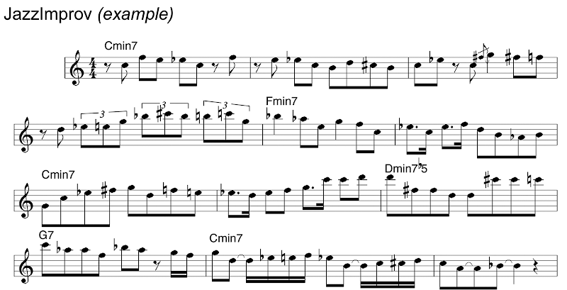
As a researcher, I was impressed with the authors' clever study design, which put 6 professional jazz pianists (one at a time, that is) into an fMRI machine with a non-ferromagnetic MIDI keyboard. They then recorded the MIDI output from this keyboard while creating simultaneous fMRI maps of brain activity. First, the musicians played a control piece: a simple one-octave C major scale in quarter notes. They were then asked to play an improvised melody, but were restricted to the use of C major scale quarter notes within the same octave. Next, they were asked to play a pre-memorized original jazz composition, in synchrony with a pre-recorded music-minus-one jazz quartet. Finally, they were allowed to cut loose and improvise freely with the pre-recorded accompaniment.

JazzImprov-journal.pone.0001679.g001-cropped.png

The authors conclude:
Our results strongly implicate a distinctive pattern of changes in prefrontal cortical activity that underlies the process of spontaneous musical composition. Our data indicate that spontaneous improvisation, independent of the degree of musical complexity, is characterized by widespread deactivation of lateral portions of the prefrontal cortex together with focal activation of medial prefrontal cortex. This unique pattern may offer insights into cognitive dissociations that may be intrinsic to the creative process: the innovative, internally motivated production of novel material (at once rule based and highly structured) that can apparently occur outside of conscious awareness and beyond volitional control.
As a radiologist, I'm delighted to see cool new uses of the machines I work with every day. When I started my career, we were happy just to be able to distinguish gray matter from white matter. It now appears that we are beginning to look at some of the nuts and bolts of creativity itself.

As a musician, a part of me will continue to nurse a small, Luddite hope that we don't discover all of the brain's secrets too quickly. After working with high tech gear all day, it's great solace to retreat to the atavistic pleasures of folk music and dance -- things I can enjoy even miles from electricity. For now, I'll continue to routinely deactivate major portions of my prefrontal cortex when I'm away from work, and let my instruments and my body move through the music on cerebellar cruise control alone.

(hat tip to Medgadget, whose post, This is Your Brain on Jazz, has a much better title than mine.)


Update, 3/5/08: This PLOS paper has just been featured in USA Today. To see what a keyboard player in an MR scanner looks like, see here.

Monday, March 3, 2008

National Museum of Health and Medicine Images Online

If you just stick to the Capitol Mall on visits to Washington D. C., you will probably never end up at the wonderful National Museum of Health and Medicine. If so, you'll end up missing out on an awesome and ginormous collection of historically and medically fascinating items. This museum was founded as the Army Medical Museum in 1862, and their collection now includes over 24 million items.
Skull radiograph above shows a 0.50 caliber bullet wound of the face. Injured while heating 0.50 caliber incendiary m.g. [?] bullet with a blowtorch while manufacturing an ash tray. Entrance just below right eyebrow. 11 days after injury, patient began hemorrhaging; blood flow so profuse it was impossible to carry out emergency procedures. Patient deceased. World War 2.


"Crossing your fingers" won't prevent venereal disease but a prophylaxis will. Venereal Disease Control Office, L.J. Stephens, Captain, Medical Corps, Jefferson Barracks, M.O.
As part of my radiology training, I got to spend 6 weeks at the AFIP course in radiologic pathology. Since the NMHM and AFIP shared the same building at the time, I spent a lot of my spare time wandering through the NMHM exhibits. Some of my favorites: bits of Abraham Lincoln's skull, President Garfield's assassin's spleen, an isolated humerus with flagrant osteomyelitis from a Civil War minnie ball wound, hand-written medical texts by Viet Cong medics, and artwork signed by a young Army physician: Captain Frank Netter, M.D.


It's great news to hear that the NMHM has been uploading their pictures to Flickr since September 2006, under a very liberal Creative Commons license. This is a great public domain resource for medical bloggers or anyone else interested in the history of medicine.


General Daniel Sickles' leg bones
, removed after the Battle of Gettysburg during the Civil War.
Check out the following links to some of their images on Flickr at:

Otisarchives1
Otisarchives2
Otisarchives3


Other excellent images can be seen online at the Otis Archives Gallery.

(via Morbid Anatomy)

Sunday, March 2, 2008

Radiology Eye for the Straight Guy

"There he is in that thicket -- his ear just twitched!"
My uncle has just described something that I cannot see at all. I look harder, but no joy.
"Look, he's standing up!"
I peer through the scope of my rifle, but still see nothing but distant thicket, about 200 - 300 yards away from us.

Finally the deer bolts from the thicket and heads away from us at Warp 2. At this point, even I can finally see the darned deer. Of course, the butt of a running whitetail deer is only slightly harder to see than a flashing strobe light. For someone who normally takes pride in having sharp vision, I am feeling pretty humble right now.

This happened years ago, just before I headed off to medical school. However, I remember it very clearly, because it was the first time I truly saw the vast difference between what can be seen by a trained eye versus an untrained eye. This was a recurring lesson in med school, whenever one of my professors would make an astute diagnosis from some minor clinical clue I had never noticed.

Nowadays, I spend my workdays interpreting medical images, usually side by side with the medical students or radiology residents I teach. I now possess the same advantage over them that my uncle had over me - decades of practice at training my eye to see tiny details in a sea of noise.

My students still remind me of what it's like to have the eye of a beginner whenever they miss something that seems now seems quite obvious to me. I then get to point out to them the bent blade of grass or the twitching ear that gives away the deer hiding in the picture. Usually, most of my students believe me, and accept this lesson with appropriate humility.


However, sometimes they don't. I was once confronted by an incredulous med student in the middle of a trauma lecture I was giving to his entire second year class. I had just suggested to them that the patient above with knee pain had a fracture of his femur. Before reading any further, take a moment and see if you can spot the abnormalities that they didn't see.
"You can't expect me to believe that you see a fracture on that film!"
Judging from the vocal borborygmi coming from all around the room, his classmates didn't believe me either. Even after I pointed out the subtle X-ray findings, they didn't seem very convinced. At the time, I was somewhat disconcerted by their distrust.

In retrospect, it's not hard to see why they were so skeptical. To them, I was a magician who had walked out on stage with a rabbit in his hand, and was now trying to convince them that I pulled it out of my hat backstage. Why should I expect the trick to work if I didn't first show them the empty hat or my empty hands?

With the benefit of hindsight, what would I say to them now? The French have a great term for this predicament : "l'esprit de l'escalier" (literally "stairway wit") -- the comeback you only think of when it is too late. Here's mine:
I don't blame you for being skeptical. Extraordinary claims require extraordinary proof, and so far, the only evidence you have that I spotted this fracture is my say-so.

Critical thinking is one of the skills we want you to learn here in medical school. I'm glad that you are using it here, and I admire your being courageous enough to voice your disbelief in front of your whole class.

So, you don't have to believe that I spotted that fracture. But please believe this: that an expert observer can spot things that are invisible to a newbie.

As you move from your classroom years to your clinical rotations, you will see this phenomenon again and again. You will see cardiologists finding heart murmurs that you can't hear. You will see surgeons palpating breast masses that you can't feel. And yes, you will see other radiologists spotting fractures that you can't see.

You will also do these things yourself, once you have enough flight time of your own.
At this point, I would again point out the findings that shout "Fracture!!" very clearly to my eye:
1. the tiny discontinuity in the femoral cortex that shouldn't be there


2. the straight line formed by a fat-fluid level in the knee joint -- a handy sign that a bone has broken, allowing marrow fat to leak into the joint


I would also tell them the story of my uncle , and what I learned about reading films from him and the deer.  Even though I didn't realize it at the time, that day long ago in West Texas may have been the first real day of my training as a radiologist.

Saturday, March 1, 2008

Escape from Paper Mountain

In the days of yore (about 5 years ago in internet years), radiologists interpreted physical films. Many of these films were large (14" x 17") , were stored in large paper envelopes, and were theoretically stored in a film library, which occupied a large chunk of every radiology department. This theoretical ideal was approached only in film libraries run by file room gnomes as fanatical as those in Gringott's Wizarding Bank. In practice, most of these libraries operated in a state of barely controlled chaos, with film folders squirreled away in doctor's offices and locker rooms throughout the medical center.

Life in the medical imaging world is much better now. In the past 5 years, a large fraction of medical imaging has gone completely digital. The images are acquired digitally and stored digitally, usually in an online digital film library called a PACS (picture achiving and communication system). File room gnomes have largely been supplanted by an equally fanatical group of IT gnomes, who keep the PACS and radiology workstations up and running.

Moving to a PACS system has solved so many of our old film-based system that most of us would never go back. Images are stored centrally where many folks can view them simultaneously. Search and retrieval of images is usually a piece of cake -- most decent PACS systems automatically find and open an appropriate comparison image right next to a new exam.


Now that this major part of my life has been enhanced by going digital, I've eager to find ways of upgrading other parts of it. Even though my office is no longer dominated by stacks of films, it continues to be crowded by paper. My file cabinets, desktop, and yes, even my floor are filled with journals, manuscripts, forms, letters, research data, receipts and lots of other crap. This paper glacier even extends an arm into my home. It was therefore with great interest that I ran across an article on the 43 Folders personal productivity site called Palimpsest: the guide to a (mostly) paperless life, by Ryan Norbauer.

This article echoed a lot of my own feelings on the tradeoffs between paper and digital:
Paper also lacks a number of the affordances of its digital counterparts: pen and paper don't offer very good full-text search, for one thing—and the spell check is even worse. But as a medium for encouraging unbounded creative thinking and planning, nothing quite beats a hunk of the old papyrus.
Another great quote:
... the key is to recognize that paper is all about process, whereas digital media are all about information and retrieval.
Bold words, but just how does one convert mounds of paper into some useful digital form? For me, so far, the answer has been the phenomenal ScanSnap S510M, by Fujitsu. This little jewel lets me drop a stack of papers into its built-in automatic document feeder, hit one button, and then ignore it while it converts the stack into a PDF file at up to 36 pages / minute in duplex mode. The ScanSnap scans both sides simultaneously in color, at an optical resolution up to 600 dpi. The scanner comes with a full copy of ABBYY FineReader, which converts the PDF's into searchable PDF's. This latter feature is the final clincher for me. In this context, searchable means that one can search for a particular word within the scanned PDF. For me, this works either while viewing a single PDF or while searching my hard drive via Spotlight.

Since the ScanSnap scans both sides of a document simultaneously, clippings often include unwanted material as part of the PDF. However, it's very easy to use Apple's awesome Preview or Adobe's Acrobat to rearrange or remove pages. A full copy of Acrobat comes bundled with the ScanSnap, by the way.

This might be a good place to mention that the ScanSnap S510M is a Mac-only device. However, Fujitsu makes a version for Windows-based PC's as well, called the ScanSnap S510. As far as I can tell, these devices are otherwise identical, other than their colors (white = Mac; black = Windows). Read into this color scheme what you will.

One last nicety: either of these devices have a footprint small enough to sit on a small corner of my desk (less than the size of a standard US piece of paper). The ScanSnap also folds up, making it easy to carry to a new location.


One final bit of the roadmap out of Paper Hell is finding the right place to store the stuff. At the moment, I've been using the iDisk on my .Mac account. After scanning in a file, I drop it onto some meaningfully-named folder on my iDisk icon, which causes the file to be synchronized automatically with Apple's servers. Files on my iDisk are then available to me at other locations and other computers from across the net, but are not accessible to the public.

Another alternative for offline storage is Amazon's new S3 web service. S3 stands for Simple Storage Service, and Amazon currently charges 15 cents / GB-month of storage used. About this, Norbauer says:
I use JungleDisk to automatically back up my "inbox" and "Archive" folders to S3 each night (not to mention iTunes and iPhoto.) Now it actually seems more dangerous to keep a single physical copy of a document than to have digital copies on my hard drive and redundantly backed up to S3. You can read here all about the baroque lengths Amazon goes to in order to safeguard your data on S3.
Both Norbauer and Gordon Meyer, of the O'Reilly Network MacDevCenter, give a number of practical tips about how they set up a personal workflow with the ScanSnap. Meyer also describes the programs he uses to wrangle his growing herd of 1,000+ PDF files, such as DevonThink Pro Office, Skim and PDFPen.

Soooooo.... how is all of this working out for me so far?

Thus far I've scanned in 218 files, which are currently backed up offsite on my iDisk account. Each file probably runs an average of 5 pages, so I'm already up to the 1,000+ page mark myself. This includes all sorts of receipts, manuals for home appliances, and lots of other papers I'd like to be able to easily find again.

This process has also been very useful in managing my son's increasing homework loads. After scanning in an assignment, a copy is e-mailed to his teachers and then stored in the iDisk archive. Another pending project -- using the ScanSnap to digitize and store a lot of his early school artwork that is currently in bulging boxes around the basement.

Finally, the ScanSnap has turned out to be a great alternative to fax for us. The ScanSnap scans faster than our home fax machine, and allows us to e-mail things out in one large PDF via our home high-speed internet connection. This also saves having to tie up the home phone line while a large stack of papers oozes slowly out of the house at fax modem speed.


I initially got a ScanSnap for home use. However, it quickly became so useful to me that I soon bought another one for my desk at work. I've still got several metric tons of paper to go, but for the first time in a very long while, I feel like I've got options other than a bulldozer and a landfill for dealing with them.